

The second session of “Cardiovascular Ultrasound Training for Small Animals” in the northern region was successfully completed!
Release time:
2022-04-19
On April 15, the “Small Animal Ultrasound Training” organized by Yik Lecture Theatre - Northern Region Online Lecture was successfully concluded. This meeting was held by our many years of rich experience in imaging, Mr. Wu as the instructor of the hands-on class, during the live communication and communication of hands-on experience, to discuss how to solve the ultrasound imaging and analysis of the difficult points, so that teachers quickly and comprehensively master the ultrasound scanning skills.

Wu Yifeng Product Technical Engineer
Engaged in ultrasound imaging technology for many years with a wealth of experience
Course content:
● Evaluation of mouse left ventricular systolic function ultrasound scanning techniques.
● Mouse aortic vascularization scanning techniques.
● Evaluation of left ventricular diastolic function in mouse ultrasound scanning techniques.
● Carotid artery vascularization in mice
● Coronary artery blood flow scanning in mice
● Mouse aortic vascularization scanning techniques.
● Evaluation of left ventricular diastolic function in mouse ultrasound scanning techniques.
● Carotid artery vascularization in mice
● Coronary artery blood flow scanning in mice

Help teachers to answer more questions in the interactive exchange on site

M-mode of left heart function in mice
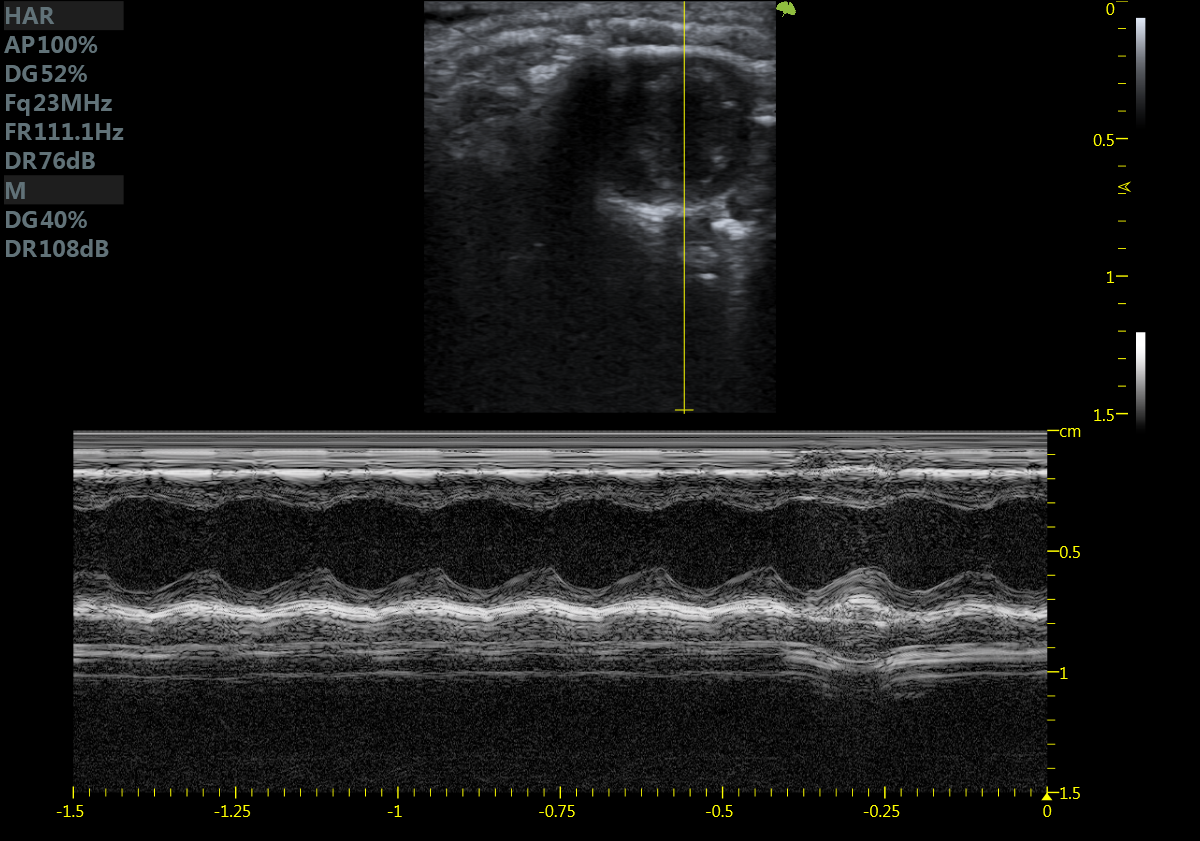
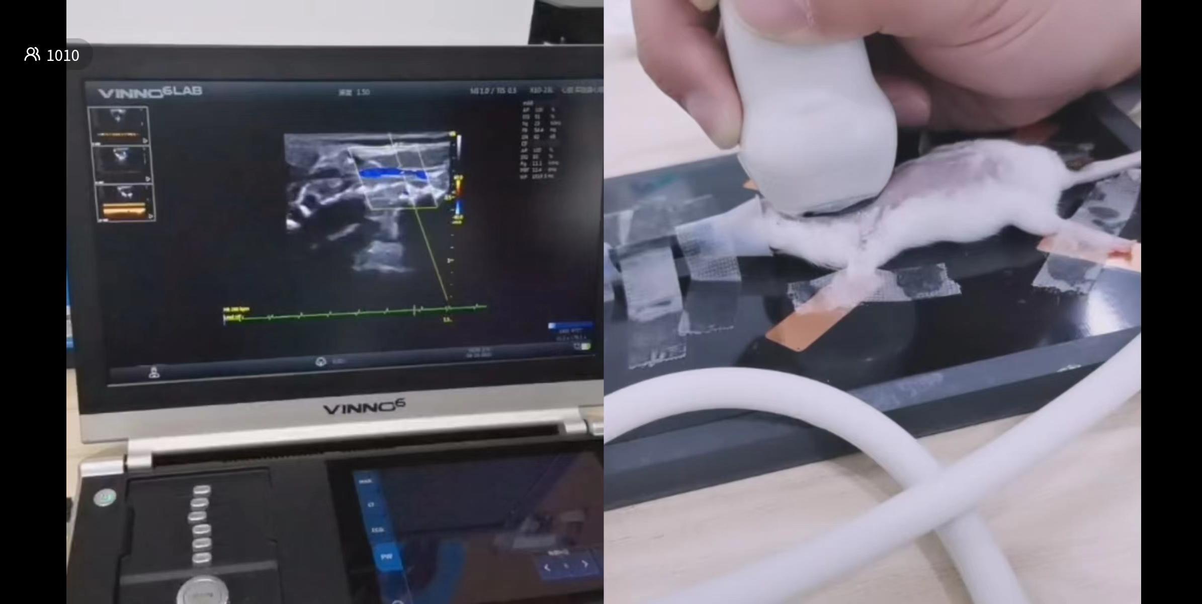
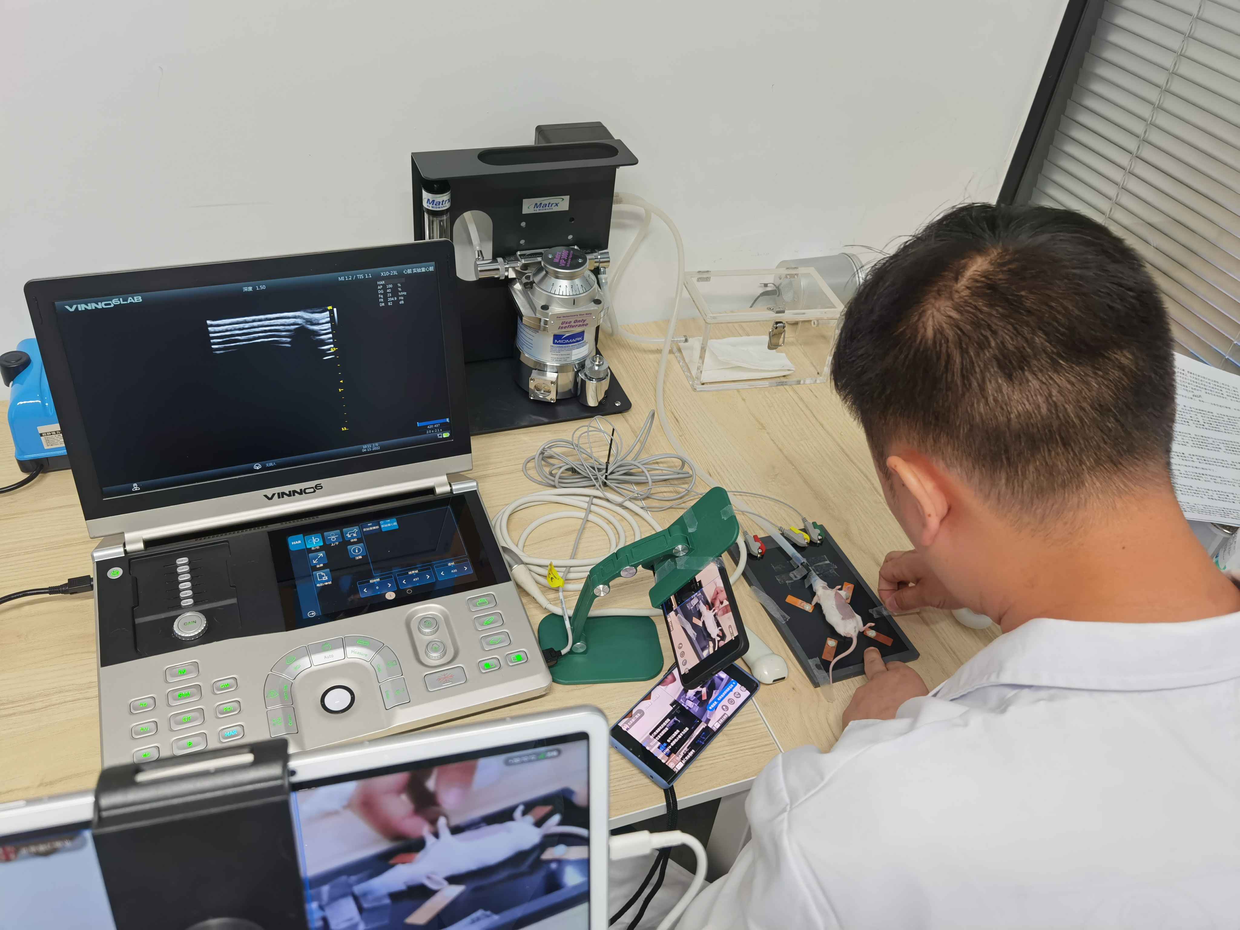
Measure the left parasternal short-axis view in mice, the ultrasound probe is placed at the left sternum, the probe marking point is directed toward the 3 o'clock position, presenting the left ventricular papillary muscle view, and click on the M-mode to get the M-mode image of the left ventricular short-axis view.
Mouse Coronary Blood Flow
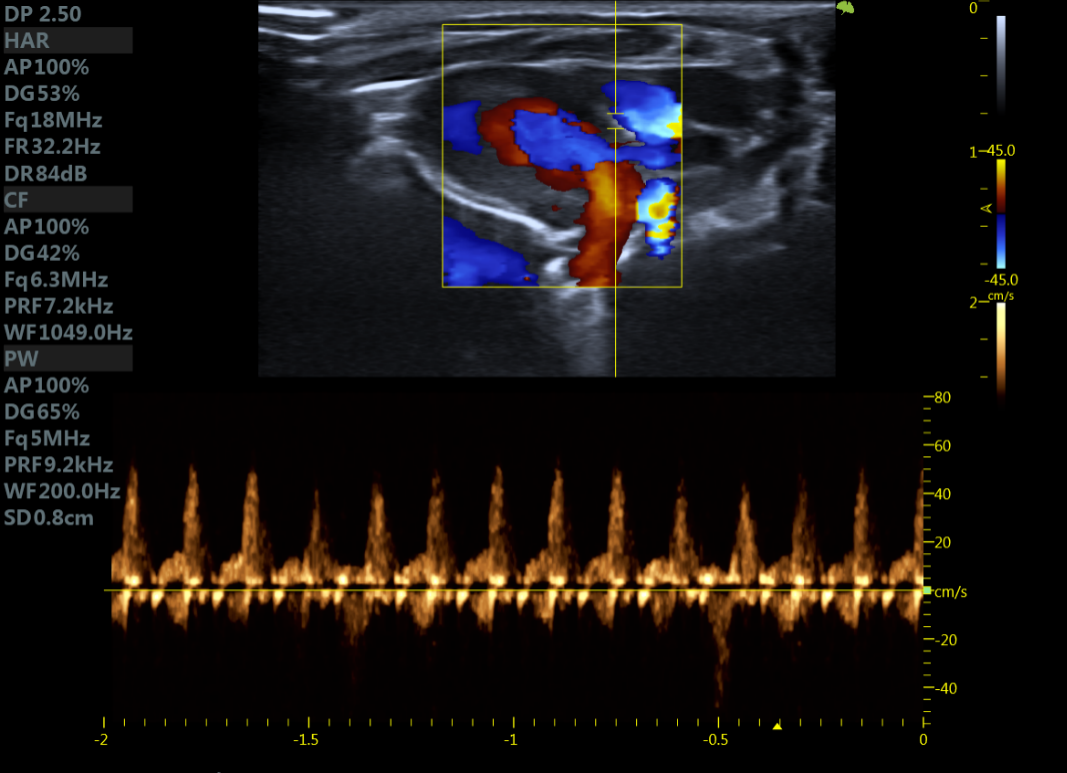
Measurement of mouse coronary blood flow, based on in the left ventricle long-axis section, turn on the blood flow function, micro-adjustment, can show a clear coronary blood flow signal, the sampling volume is placed at the blood flow signal, you can measure the blood flow spectrum of the coronary artery.
Mouse aortic arch
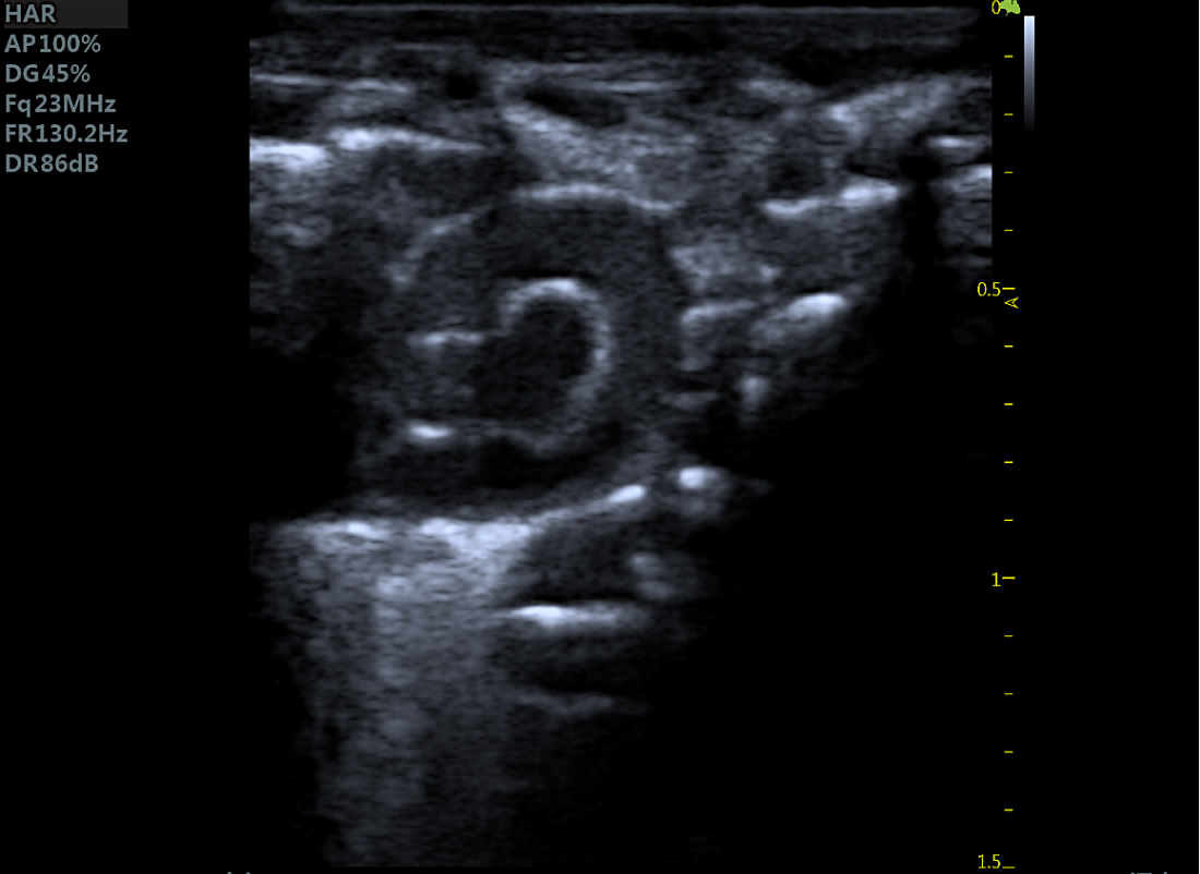
The ultrasound probe is positioned with the marker point facing the head, and the probe beam is directed from the right side of the chest. After fine adjustment, the aortic view can be found, and the ascending aorta, aortic arch, descending aorta, and the three branches can be seen.
We also have a raffle for this training:
First Prize: Philips Bluetooth Headset
Second Prize: Xiaodu Smart Speaker
Third prize: Nepal small cooking pot
First Prize: Philips Bluetooth Headset
Second Prize: Xiaodu Smart Speaker
Third prize: Nepal small cooking pot


Congratulations to the teachers who won the prizes!
Through this online learning, I believe that teachers have a good understanding of the difficult to sweep the cut surface, but also hope that in each training session can help more teachers to answer professional questions in the experiment, for your experiments to solve the problem.
▼ We will continue to conduct the “Benefit Lecture” live class, and look forward to seeing you at the next lecture!
Related News


